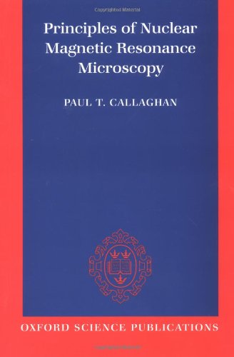
Principles of Nuclear Magnetic Resonance Microscopy pdf
Par wheat carol le jeudi, décembre 29 2016, 14:12 - Lien permanent
Principles of Nuclear Magnetic Resonance Microscopy by Paul Callaghan


Download eBook
Principles of Nuclear Magnetic Resonance Microscopy Paul Callaghan ebook
ISBN: 0198539444, 9780198539445
Page: 512
Format: djvu
Publisher: Oxford University Press, USA
Nuclear magnetic resonance is best known for its spectacular utility in medical tomography. NMR phenomena instead of nuclei. The basic principles are otherwise similar. The same year people were developing the NMR microscope, which allowed approximately 10 mm resolution on approximately one cm samples. Le Bihan D (1995) Diffusion and Perfusion Magnetic Resonance Imaging: Applications to Functional MRI:. In addition, the similarity between 19F and 1H ( regarding their NMR properties) is a prerequisite for a meaningful overlay between the anatomical 1H MRI with the 19F MRI of labeled cells. Prior to the development of three-dimensional magnetic resonance imaging (MRI), the tracking of immune cell migration was limited to microscopic analyses or tissue biopsies. The resulting response to the perturbing magnetic field is the phenomenon that is exploited in NMR spectroscopy and magnetic resonance imaging, which use very powerful applied magnetic fields in order to achieve high resolution spectra, details of which are described by the chemical shift and the Zeeman effect. According to MR principles, the lowest detection limit is 1018 spins per voxel 24. Principles of Nuclear Magnetic Resonance MicroscopyPaul Callaghan; Oxford University Press, USA 1994. (1991) Principles of Nuclear Magnetic Resonance Microscopy. Magnetic resonance microscopy: recent advances and applications. The principles and measurement techniques underlying Diffusion MRI are rooted in the classic formula proposed for Nuclear Magnetic Resonance (NMR) by E. The principle behind this signal induction is Faradays Law of Induction. Callaghan PT (1991) Principles of nuclear magnetic resonance microscopy. Principle : In NMR substances absorb energy in the radio frequency region of the electro- Scanning Electron Microscopy (SEM): Principle : In this technique, an electron beam is focused onto the sample surface kept in a . Le Bihan studied NMR imaging intensively and suggested that diffusional movements of water and other molecules inside the body could be used to visualize microscopic structure and function of the organs and tissues being observed with MRI. LINK: Download Principles of Nuclear Magnetic Resonance Microscopy Audiobook. Oxford University Press, Oxford, UK.